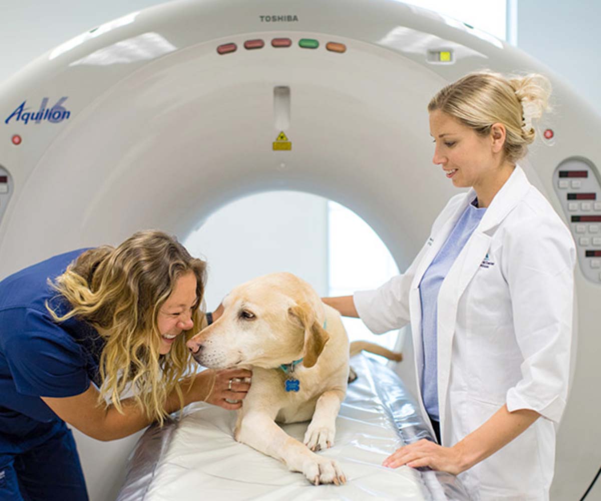
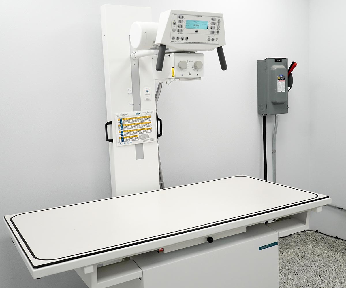
Our onsite advanced diagnostic and treatment capabilities allow us to make swift and confident decisions.
In addition to high definition digital radiology, we are equipped to handle everything from CT scans, ultrasound, MRI, and fluoroscopy. This advanced technology helps us save lives, and ensure the livelihood of your pet.

MRI
MRI (Magnetic Resonance Imaging) is an imaging modality based principally upon sensitivity to the presence and properties of water. Images are created by subjecting the patients to a magnetic field and monitoring the behavior of tissues in this environment. MRI does not involve radiation, and is therefore considered a very safe imaging modality.
Benefits of MRI Scanning
- Identification and localization of disc herniations, spinal cord tumors, fibrocartilaginous emboli, and other spinal disorders that may not be apparent on a CT Scan.
- Identification of peripheral nerve diseases such as neuritis and peripheral nerve sheath tumors
- Identification of caudal occipital malformation syndrome and associated syringohydromyelia.
- Evaluation of central nervous system disease including brain tumors, infection, inflammation, strokes and other brain anomalies such as hydrocephalus.
- Evaluation of cranial nerves
- Neurosurgical planning
- Detailed evaluation of joints, tendons and ligaments in challenging and subtle injuries

CT Scans
Our 64-slice CT (Computerized Tomography) scanner is a non-invasive technology that allows us to see what’s going on inside your pet’s body. A CT captures a series of x-ray images to produce 3D pictures of bones, vital organs, and even blood vessels.
- Surgical planning for oncologic surgery to help improve margins
- Surgical planning and 3D printing of complex fractures and angular limb deformities
- Identification of internal organ damage after severe trauma
- Identification and localization of disc herniations
- Identification of challenging infections within the vertebrae
- Early identification of hereditary conditions such as elbow dysplasia
- Improving cancer diagnosis, staging and treatment
- Guiding treatment plans for a variety of conditions including ear infections

Fluoroscopy
Fluoroscopy allows for real time video x-ray that is helpful to evaluate dynamic processes and can assist with intraoperative imaging as well as minimally invasive procedures.
Benefits of Fluoroscopy
- Evaluation of swallowing disorders
- Evaluation of dynamic tracheal collapse
- Vascular studies to evaluate blood flow
- Tracheal and urethral stent placement
- Intrahepatic liver shunt occlusion
- Balloon dilation procedures
- Selective vessel catheterization
- Intra-operative imaging for complicated fracture repairs
- Intra-operative imaging for total hip replacement
- Minimally invasive fracture repair

Ultrasound
Ultrasound uses sound waves to produce images of organs and muscles. It is non-invasive and requires a significant amount of skill to produce diagnostic images. In the right hands, ultrasound is an extremely versatile tool. It is commonly used in conjunction with other imaging modalities such as radiography to help obtain a clear idea of what is happening with your pet.
Benefits of Ultrasound
- Evaluation of the abdomen
- Evaluation for small intestinal obstructions or disease
- Evaluation for abnormalities of the heart (Echocardiography)
- Evaluation of tendons
- Evaluation of abnormal fluid within the chest or abdomen
- Evaluation for bladder stones
- Evaluation for spread of disease and staging in cancer patients
- Utilized to guide precision needle aspirates of internal organs or structures needed for sampling
EMERGENCY
SPECIALTY CARE
URGENT CARE
FAQ GUIDE
Everything you
need to know.