Advancing Orthopedic and Neurologic Surgical Care.
Our team of exceptional surgeons, nurses, and rehabilitation experts, is specifically focused on the needs of our orthopedic and spinal pet patient population. We work together to deliver the ideal solution for your pet regardless of complexity. Also integrated within this center is rehabilitation therapy which allows us to improve upon our outcomes with patient specific treatment plans.
Bone, Joint, & Spine Center
62889 NE Oxford Ct.
Bend, Oregon 97701
541-209-6960
Directions
Innovative Care
Our expert staff has a passion for pushing the boundaries and working towards outcomes not attainable anywhere else. Our team is tasked with always finding the best treatment option possible and thus constantly pushes itself to learn, innovate, and improve.
Orthopedic Surgery
Orthopedic surgery is a broad title that involves operating on the bones and joints of animals. This can include traumatic injuries such as fractures and ligament injuries all the way to congenital malformations like elbow or hip dysplasia.
Tibial Plateau Leveling Osteotomy
A tibial-plateau-leveling-osteotomy (TPLO) is a common orthopedic procedure to repair a torn cranial cruciate ligament (CCL) in dogs. This procedure is considered the gold standard by most board certified surgeons and requires a substantial amount of training, such as would be obtained during the course of a residency program, and experience to perform well. The CCL is the primary weight-bearing ligament in a dog’s knee, making it susceptible to injury. In fact, injury to this ligament is the most common orthopedic injury in dogs. There are multiple surgical approaches to correct a cruciate ligament tear. Having an experienced board-certified veterinary surgeon on your team is important to help minimize the likelihood of a complication.
Patellar luxation
Patellar luxation is typically a congenital malformation of the knee joint that leads to abnormal tracking of the knee cap (patella) along the femur. This abnormality can lead to an intermittent dislocation of the patella out of alignment which causes discomfort, pain, an abnormal gait, and degenerative joint disease.
Arthroscopy is a minimally invasive procedure used to diagnose and treat many joint problems, such as elbow dysplasia. A narrow tube attached to a video camera is inserted through a small incision. The camera feed is transmitted to a video monitor, allowing our surgeon to have a magnified, high definition view of the joint. Small instruments can then be used to treat your pet through the same small incision without a need for an invasive open surgery site. Most pets are walking on their operated limb the same day.
Fractures that cannot heal by wearing a cast will require a surgical approach for stabilization. Fracture repairs can be very complex, and may involve the use of screws, pins, and bone plates to ensure that the bone heals appropriately.
Pain control is a priority when a patient is seen for evaluation of a fracture. Diagnostic imaging is crucial for making an appropriate plan for repair. Many fractures can be properly evaluated through routine x-rays, while some may require a CT scan so that a 3D image can be made and allow for appropriate surgical planning. If needed, we will 3D Print fractures to better understand how to fix them. Special consideration must be made for fractures involving the growth plates in a young animal.
We are the first hospital in the region to start a program of this caliber. Canine Total Hip Replacements have been performed successfully in dogs for decades. With advances in technology, you can expect an approximate 90-95% success rate, which results in an excellent quality of life and the ideal solution for dogs affected with hip dysplasia.
Hip replacements can be performed as early as one year of age, simply because dogs don’t live as long as humans. Several other surgical interventions that your pet may be a candidate for include: triple pelvic osteotomy, juvenile pubic symphysiodesis, or femoral head and neck ostectomy. If your pet is diagnosed with hip dysplasia, a consultation with a boarded veterinary surgeon to discuss the pros and cons of treatment strategies is recommended right away. Some of the surgical options are time sensitive, and the progression of hip dysplasia may render the procedure suboptimal.
Neurologic Surgery
Neurologic surgery can involve both the spine and the brain of pets. Spinal surgery can be indicated for a multitude of reasons including herniated discs, trauma and cancer. Brain surgery is very specialized and utilizes very careful planning with advanced imaging such as an MRI or a CT scan prior to the procedure. A common indication for brain surgery would be to remove a tumor growing on the skull or brain of a pet. Depending on the location and type of tumor, the prognosis for brain surgery can be good.
If your dog has a slipped or herniated disc, a decompressive surgery such as a hemilaminectomy or ventral slot may be recommended by your family veterinarian. Because this operation deals with the area surrounding the spinal cord, only a surgeon specially trained in neurosurgery should perform this procedure. All of the surgeons at VRCCO are trained in neurosurgery.
The goal of the procedure is to remove the ruptured disc material to alleviate compression on the spine. Prior to the procedure, your pet will undergo a CT scan or MRI with contrast to appropriately visualize the affected disc. This allows us to perform surgery at the correct location.
This surgery may also be recommended for severe or recurrent cases of Intervertebral disc disease (IVDD). IVDD is a common ailment affecting dogs, especially breeds that have long backs, like dachshunds and other popular breeds such as french bulldogs. In general, rehabilitation therapy is highly beneficial for patients undergoing spinal surgery.
Some dogs have dynamic spinal instability which leads to chronic discomfort or neurologic decline as the spinal cord and nerves exiting the spinal cord become compressed or traumatized. Common version of dynamic spinal disease include cervical spondylomyelopathy (“Wobbler’s disease”) and lumbosacral disease. These conditions may be well managed with medical management and rehabilitation therapy however some require surgical intervention. It is important to have an accurate diagnosis and accurate diagnostic imaging to make these decisions.

Advanced Imaging
We utilize a variety of diagnostic tests to evaluate a pet thoroughly, make an accurate diagnosis and offer the best recommendations for treatment. In many cases, survey radiographs (X-ray) are not sensitive enough to give enough information for some specific disease conditions. When we need more accurate diagnostics we will utilize modalities such as fluoroscopy, computed tomography (CT), or MRI.
Magnetic resonance imaging (MRI) is a super-specialized imaging technique used to evaluate delicate soft tissue structures such as the brain, spinal cord, nerves, tendons and ligaments. In veterinary medicine, it is used most often for the evaluation of neurologic conditions such as herniated disc disease, brain tumors or inflammatory brain or spinal disease. MRI also has specialized utility for complex ligament injuries like shoulder tendinopathies.
CT is a test performed that gives us cross sectional visualization of the body that is significantly more accurate than plain radiography. The CT can be processed into three-dimensional images to give the doctor an accurate evaluation of bones, joints, soft tissues, blood vessels and even the spine. CT is also utilized for very fine- or hard to look at regions like the nasal cavity, inner ear and sinuses.
This test utilizes x-ray in a real time movie format. This is important for dynamic conditions that are not always caught on a single x-ray. Examples of where this is helpful is with evaluating how animals swallow or breathe and how blood vessels flow. Fluoroscopy is also utilized intraoperatively during complex orthopedic repairs to guide and evaluate implant placement and bone alignment without waiting until the procedure is finished to double check our work. This allows us to be as accurate as possible during surgery.

Rehabilitation Therapy
Rehabilitation therapy is a rapidly growing space within veterinary medicine. As we learn and understand dogs and cats better we have come to realize that rehabilitation therapy is incredibly important. It has a place not only in the perioperative period but also in helping older pets maintain strength, stamina, and reduce pain.
Rehabilitation therapy utilizes multiple modalities and exercises to help treat musculoskeletal and neurologic conditions that are ailing your pet. Rehabilitation therapy is sometimes the sole treatment needed to improve your pet’s condition. In other instances, it is utilized in concert with surgical treatment to achieve successful outcomes. Rehabilitation therapy modalities can include underwater treadmill therapy, cold laser therapy, shockwave ultrasound therapy, specific stance exercises and acupuncture. Your rehabilitation therapy team will work alongside board certified surgeons to evaluate your pet’s specific needs and create a customized regimen to ensure the best possible outcome. All staff in the Rehabilitation therapy department have undergone additional training and/or certification specific to veterinary rehabilitation therapy to ensure we are doing our best for your pet.
WHAT TO EXPECT
Bone, Joint & Spine Center

Each patient will undergo preoperative diagnostics specific to the procedure being performed. The most common diagnostics are blood work, x-rays, abdominal ultrasonography or CT scanning. These diagnostics allow the surgical team to make an individualized surgical plan for your pet. Your pet will receive appropriate pain control before, during, and after surgery. Click here to learn more about pain management.
Prior to surgery, an IV catheter and endotracheal tube will be placed, IV fluids started, and anesthesia will begin. The surgical area is then clipped and cleaned. While under anesthesia, your pet will be closely monitored by an experienced veterinary nurse anesthetist who will work with the team to ensure a comfortable and uneventful anesthetic event..
We use advanced equipment to monitor heart rate, respiratory rate, body temperature, blood pressure, heart rhythm, carbon dioxide levels, and sometimes even blood gasses. Your pet will be closely monitored until they are fully recovered from the anesthetic event. Additional precautions are always taken when performing anesthesia on inherently higher risk patients such as brachycephalic breeds.
Appropriate pain management is critical before, during and after surgery. We provide a variety of progressive anesthetic and pain management techniques, known as “multimodal anesthesia,” including:
• Continuous infusions of pain medications
• Local nerve blocks
• Epidurals
• Oral medications
• Long acting local anesthesia
All surgical patients will receive overnight, post-operative monitoring by skilled veterinary staff with doctors in the hospital 24 hours a day. Most patients are ready to go home the next day.
Two week rechecks will be performed by our Rehabilitation therapy doctor to further discuss the rehabilitation process and present the ideal therapy plan for your pet to help the recovery process be as successful as possible. It is here that our doctors may recommend rehabilitation therapy sessions at our bone, joint, and spine center. Most patients will eventually come back at the 6-8 week mark for an additional progress check with your surgeon. If your pet underwent a surgery that required implants, radiographs will likelybe taken at this time to evaluate for bone healing.
There are situations in which rehabilitation therapy is the first treatment of choice or the only treatment. In this circumstance you will be scheduled to see our rehabilitation therapy doctor. After a thorough evaluation, diagnostics, and understanding of your pet’s condition and all available treatment options, we will work toward designing a program to address your pet’s underlying condition. At the Bone, Joint, and Spine center you do not have one doctor but instead a team of doctors constantly collaborating to create the best treatment plan for your pet.
A treatment plan will be provided for any recommended diagnostics and/or treatments. An initial payment will be required for any procedure, surgery, or diagnostic work-up over $600. We gladly partner with CareCredit and Scratchpay to offer financing services to our clients. Please ask our team for more details. We are also happy to assist with submission of pet insurance claims to your pet’s insurance provider. We provide a 10% discount for pet parents in military service.
We accept cash, VISA, Mastercard, Discover, CareCredit and Scratchpay. Personal checks and American Express are not accepted.
Bone, Joint & Spine Center Team
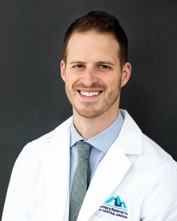
Dr. Mauricio Dujowich
DVM, DACVS SURGERY, CCRT
Dr. Dujowich’s passion for improving patient outcomes has led to becoming certified in canine rehabilitation therapy and creating the Bone, Joint, and Spine center within VRCCO. The goal for the center is to have a much more intentional approach to orthopedics, joint replacement, spinal surgery, and rehabilitation therapy so we can maximize success.
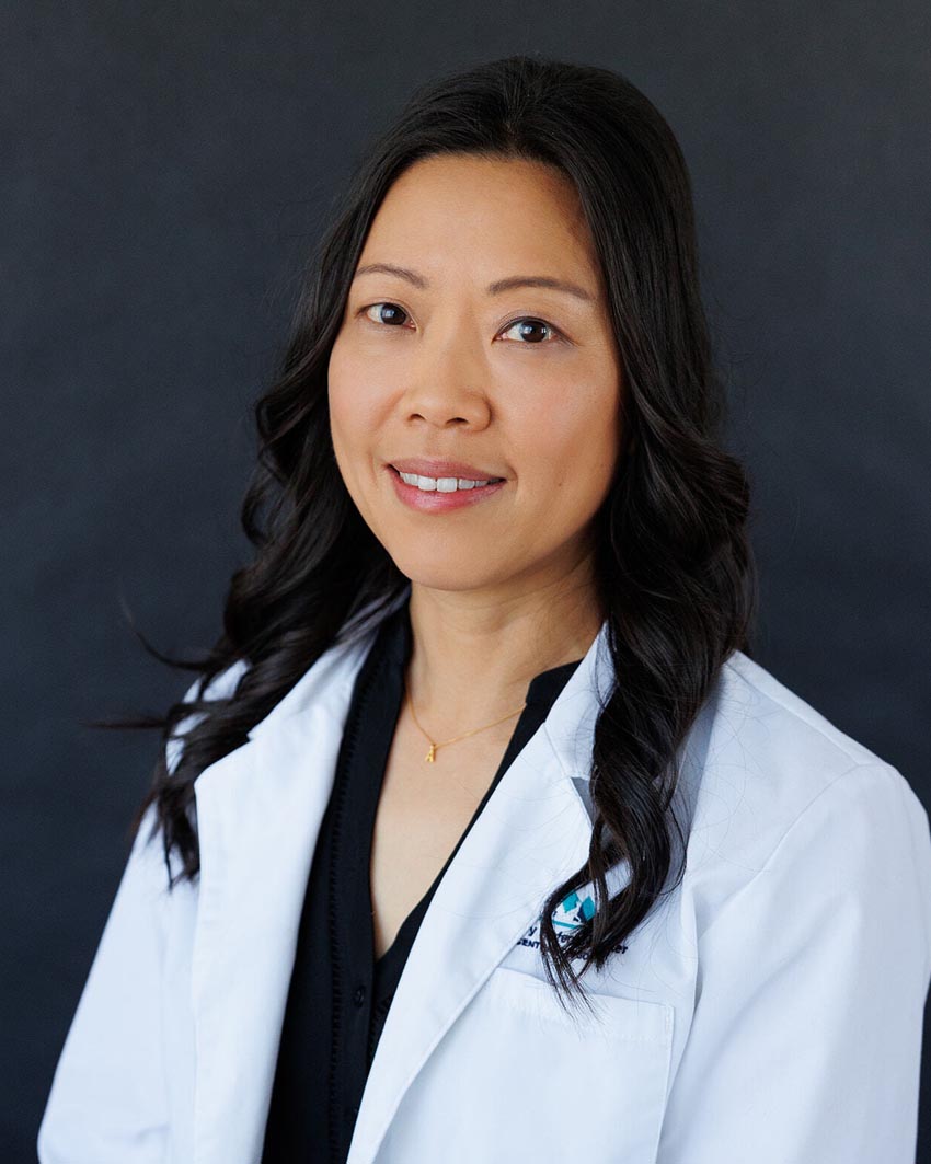
Dr. Annie Chen-Allen
DVM, MS, Dip ACVIM NEUROLOGY
Dr. Chen-Allen is a boarded neurologist with a special interest in neurosurgery. She performs spinal surgeries commonly for intervertebral disk disease, spinal fractures, spinal tumors, wobbler syndrome and arachnoid diverticulum. Common procedures performed include hemilaminectomy, ventral slot, vertebral distraction and stabilization, and spinal reconstructive surgeries. She has over 20 years of neurosurgical experience, and her team is dedicated to providing your pet with the best diagnostics and treatment options available in our state-of-the-art facility.
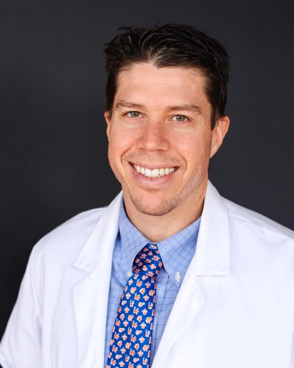
Dr. Stephen Stockdale
DVM, DACVS SURGERY
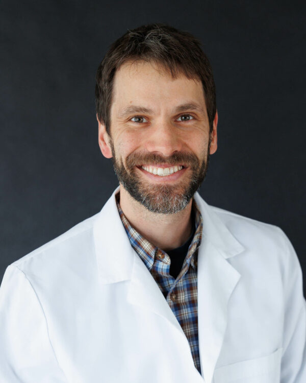
Dr. Jim Perry
DVM, PHD, DACVIM-O, DACVS SURGERY
Dr. Colin Taylor
DVM, MSC, BS, DACVS SURGERY
Dr. Ali Majer
DVM, SURGERY RESIDENT
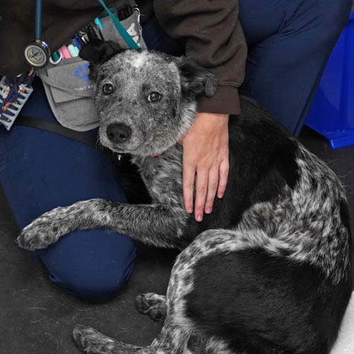
WATCH Meet Lloyd, A Success Story
Pet Hero Story
LLOYD DEFIES THE ODDS
“Lloyd got hit by a fast-moving Chevy and our local vet didn’t know everything that was wrong with him so they set up an appointment with VRCCO. He went through about five surgeries in six days, we did one at a time to see how everything was going to go, everything went well so we kept doing it. As far as I am concerned they did a wonderful job. My dogs are like my kids, they go everywhere with me, so it was well worth it.”
– Anthony Hill
The Bone, Joint & Spine Center is located at our Veterinary Emergency and Specialty Hospital in Bend. Let us know you’re coming by calling 541-209-6960 or email us at info@vrcvet.com
Bone, Joint, & Spine Center
62889 NE Oxford Ct.
Bend, Oregon 97701
541.209.6960